Pediatric qEEG fundamentals · Lesson 1
Read the background
before the trend.
Follow a child's EEG from wakefulness through drowsiness and sleep. Connect the raw patterns to their qEEG frequencies, then identify the transitions yourself.
Part 1 of 5
Start with awake backgrounds
By the end, you can…
- Compare awake backgrounds across childhood.
- Recognize drowsiness, sleep spindles and diffuse slowing in sleep.
- Locate PDR, spindle and muscle activity on qEEG, then confirm it in raw EEG.
- Mark the transition to drowsiness and the first clear evidence of N2 sleep.
Hz means cycles per second. A channel shows the voltage difference between two electrodes.
With eyes closed, look for the posterior dominant rhythm (PDR) in the derivations ending in O1 and O2. Compare its frequency, symmetry and organization with the lower-voltage activity anteriorly.
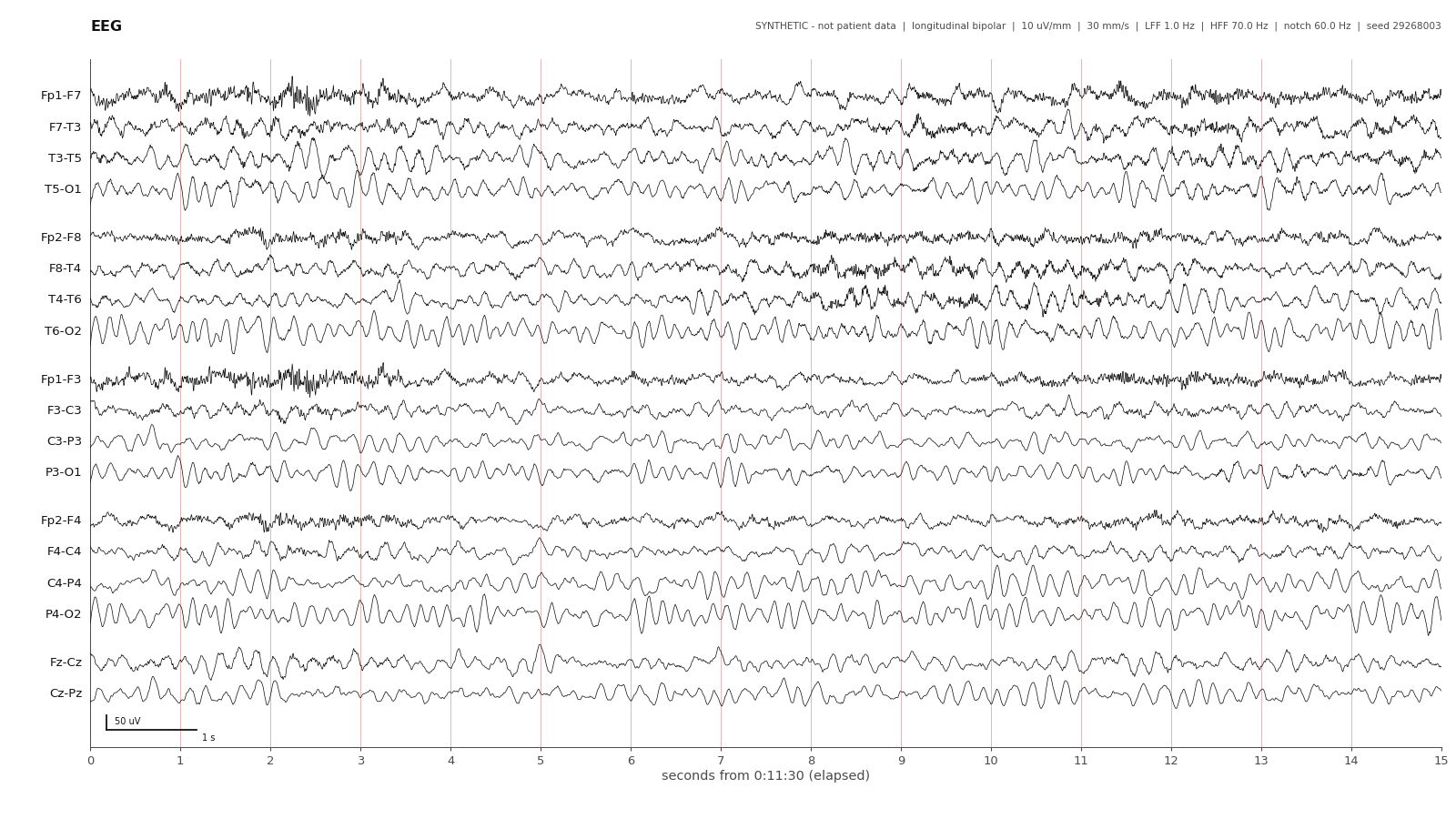
Calibration bar: 50 µV vertical · 1 second horizontal. Grid lines: 1 second.
Two-year-old awake, eyes closed: a 6-7 Hz posterior rhythm in the occipital and parietal chains with intermixed theta; lower-voltage faster activity in the frontal derivations.
Explore this recording ↗ (opens in a new tab)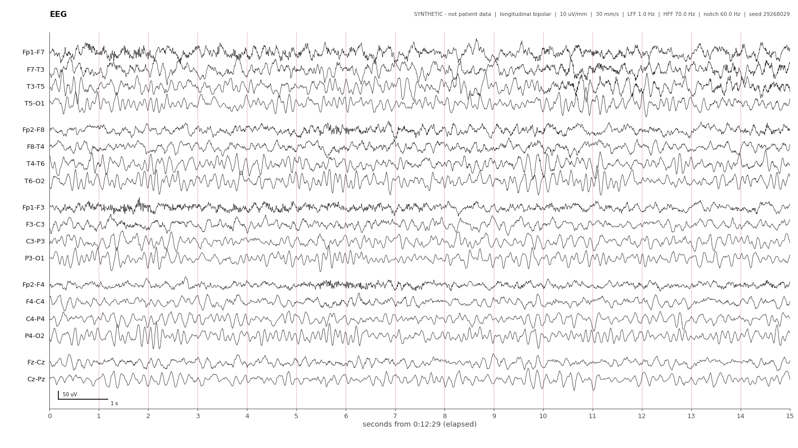
Calibration bar: 50 µV vertical · 1 second horizontal. Grid lines: 1 second.
Eight-year-old awake, eyes closed: symmetric 8–9 Hz posterior dominant rhythm, with lower-voltage frontal activity and a brief burst of temporal muscle artifact.
Explore this recording ↗ (opens in a new tab)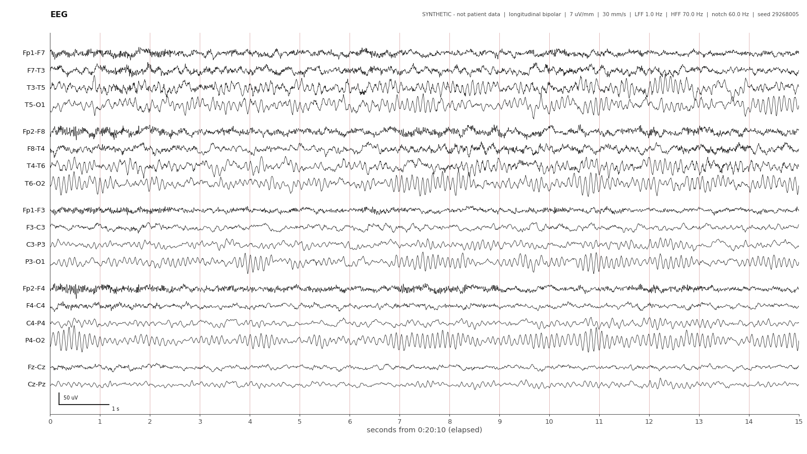
Calibration bar: 50 µV vertical · 1 second horizontal. Grid lines: 1 second.
Adolescent awake, eyes closed: a 9.5–10 Hz posterior dominant rhythm, waxing and waning in the posterior derivations, with a clear anterior-posterior gradient.
Explore this recording ↗ (opens in a new tab)These examples progress from a 6–7 Hz PDR to 8–9 Hz and then 9.5–10 Hz. Describe what you see in the context of age.
These frequencies describe the examples, not a complete table of age cutoffs. An eyes-closed excerpt alone does not establish reactivity to eye opening.
Teaching sources and example provenance
The raw examples are accepted PedQuEST gallery material. The trend figures are newly derived from the existing child recording. All recordings are synthetic.
- Toddler (2 years), awake
- Child (8 years), eyes closed: posterior dominant rhythm
- Adolescent, awake
- Drowsiness
- N2: sleep spindles
- N3: slow-wave sleep
- AES EEG atlas: The Normal EEG
- Learning EEG: Normal Asleep EEG
- Learning EEG: The Pediatric EEG
Links open in a new tab. The lesson's N2 target is the first clear spindle, distinct from the earlier drowsiness/N1 transition.